The discomfort of knowing
MRI reveals body wear and tear in extraordinary detail. What should we do with that knowledge?
By Professor Alister Hart
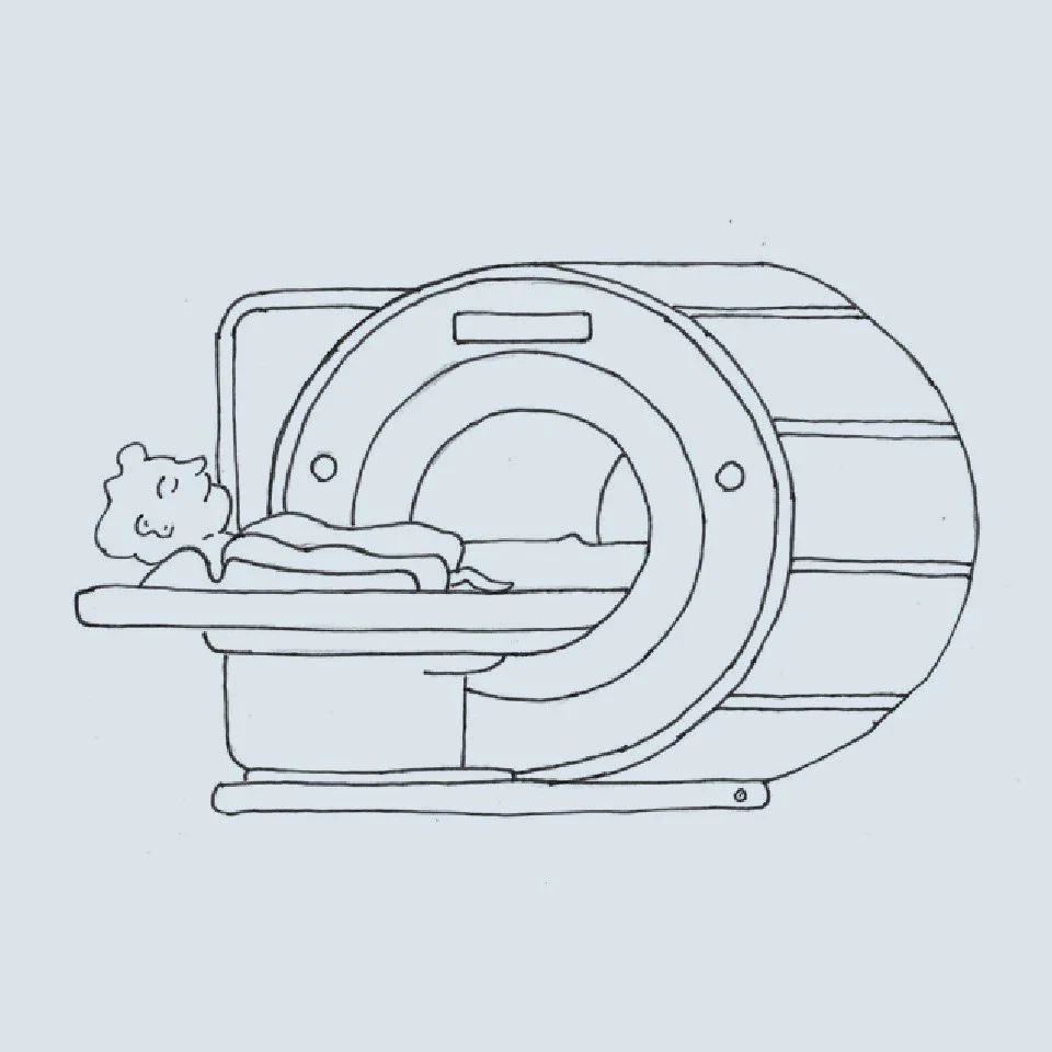
Behind the Georgian façades of Harley Street, London’s historic home of private medicine, machines are hammering. Mechanical clattering, knocking, and buzzing escape through basement windows all day. Inside, someone lies still while an MRI scanner maps the intricate structures beneath their skin.
MRI is becoming an increasingly familiar part of medical care. NHS England’s imaging dataset recorded 4.9 million MRI examinations in England in 2024–25, an increase of around 10% in a single year. More scans mean more opportunities to catch serious problems, but also a deluge of unexpected findings to deal with. MRI images reveal details of wear and tear that we weren’t looking for and may struggle to put out of our minds. My research with healthy volunteers has taught me how much hangs on the way we explain the image.
You felt great until you saw the scan.
Yesterday you were planning your next run. Now you’re unsure about walking to the shops. It turns out you have a meniscal tear, a worn disc and a problem with your hip cartilage. Labels such as “tear” and “degeneration” are difficult to take lightly when they have your name attached.
Neurosurgeon Richard Hayward gave this predicament an unfortunate name: VOMIT, or “victim of modern imaging technology”. His 2003 article in the British Medical Journal described how unexpected findings could trigger anxiety, further referrals and potentially unnecessary treatment. His warning now applies to a much larger number of consultations. Every unexpected finding brings another decision about whether to investigate, treat or reassure.
Jerome K. Jerome would have recognised the symptoms. His narrator in Three Men in a Boat visits the British Museum to read about a minor ailment. He drifts into other diseases, works through them alphabetically and becomes convinced he has almost everything. The sole exception is housemaid’s knee, an omission he finds rather insulting.
“I had walked into that reading-room a happy, healthy man. I crawled out a decrepit wreck.”
— Jerome K. Jerome, Three Men in a Boat
MRI gives this familiar anxiety something persuasive to work with: a picture of what’s happening inside your own body. Whole-body screening extends this dilemma to people seeking a scan without a particular symptom to investigate. That makes it especially important to establish whether finding more will actually improve their health.
The picture looks worse than you
Symptoms prompt most clinical scans. My research has allowed me to look at people who aren’t seeking treatment. We’ve undertaken around 1,000 MRI scans of spines, hips and knees in runners and cyclists, top athletes and couch potatoes, outdoor enthusiasts, whitewater kayakers and even Nepalese mountain porters. We wanted to understand how movement affects joints.
We also discovered how often healthy people have scans that look anything but healthy. These studies give us a useful reference point as more patients arrive with reports listing tears, degeneration and other abnormalities. Our London Marathon study found meniscal tears in just over a third of the knees of runners who completed the race and returned for follow-up scans. A meniscus helps distribute load across the knee, so a tear sounds like a good reason to reconsider several months of running. Yet these runners completed the marathon, and the follow-up scans showed no significant overall worsening of meniscal findings.
Our wider research also found tears in the cartilage around the hip socket and changes in spinal discs in people who had no pain in those areas. Some were running marathons with findings that might have made them hesitate to exercise had they known about them. These findings also occur in people without pain, so discovering one in a painful joint doesn’t necessarily explain why it hurts.
A tear is not a verdict
Imagine your knee hurts and an MRI shows a meniscal tear. You have a keyhole operation, followed by physiotherapy, and a few months later you feel better. Did the surgeon fix the problem, or does the physiotherapy deserve more of the credit?
Trials have compared surgery with physiotherapy for meniscal tears associated with ageing and wear, questioning the additional benefit of an operation for many patients. Another trial compared removal of damaged meniscal tissue with a sham procedure, in which the tissue was left in place, and found no better outcomes from removing it.
Our volunteer studies offer a complementary insight: a tear can be present without causing pain or limiting activity. Seeing one on a scan is therefore not, by itself, a reason to operate.
I assess strength, movement and how symptoms developed when examining a painful knee. Weakness or poor coordination can alter the forces around a joint. A visible tear gives us something definite to blame and may distract us from these less easily pictured explanations. I want to be confident that treating it will help before recommending an operation.
The case for knowing
MRI can reveal life-changing discoveries among the routine signs of wear and tear. A timely scan gives us a chance to act before the consequences become serious.
Stress fractures are a good example. Repeated loading can exceed a bone’s capacity to repair itself. MRI can reveal the resulting injury early enough for us to interrupt training, protect the bone and investigate why it has become vulnerable.
The stakes are particularly high around the hip. We see runners whose injuries require urgent treatment and sometimes surgery. Earlier detection may prevent an injury from reaching that stage.
A chance discovery can also be crucial. “Incidental” describes how we found something; it says nothing about how valuable that knowledge might prove. The art is in recognising which findings we can safely live with and which call for action to prevent serious harm.
More, faster.
AI is helping scanners produce more information in less time. AI-assisted protocols have substantially shortened examinations at my hospital, creating capacity for more scans. Automated analysis is also expanding what we can measure in each image.
Measuring individual muscle volumes used to mean tracing their outlines through a series of images. Software can now complete days of work in minutes.
A patient recovering from a serious injury might want to know whether their muscle wasting is progressing. Looking at a limb or measuring its circumference gives a broad impression. MRI lets us measure individual muscles and track changes that could guide treatment.
The comfort of understanding
I find these developments enormously encouraging. As a surgeon and a runner, I want to understand how our bodies respond to exercise and how we can help them recover. These tools let us investigate questions that were once impractical to pursue.
I keep coming back to our marathon volunteers. Their scans might have made them think twice about training. Four months later, their knees carried them across the finish line. As scans become more commonplace, more people will need help understanding findings like theirs. I want MRI to help them discover what their bodies can do, with the confidence that comes from understanding what we see.